What is High-Content Microscopy?
by Zacharia Nyambega
Learning Objectives
- Define what High-Content Microscopy is and list some of the High-Content Microscopy technologies available.
- Evaluate the differences between high content microscopy and traditional microscopy.
- Discuss how to use high content microscopy and state some of the other applications of high content microscopy
- List the advantages and disadvantages of High-Content Microscopy
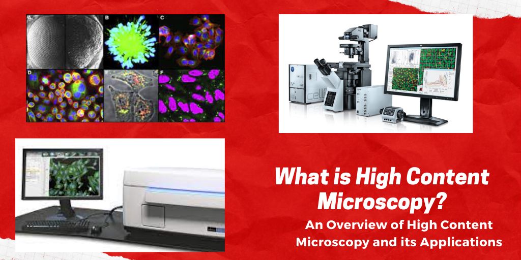
Graphical Abstract
Introduction
In the present era, scientific research is characterized by the implementation of high content-based technologies. High Content Microscopy (HCM) is a technique used in biological research that applies fluorescent detection as well as multiparameter algorithms to capture and quantify the interaction of cells. Cellular, as well as subcellular phenotypes, are visualized with the help of fluorescent tags. HCM allows parallel monitoring of various cell phenotypes (Boutros et al., 2015). This allows researchers access to various data points per cell. Examples of HCM technologies include KineticScan, BD pathway HT, ImageXpress ultra, cellWoRx, and IN Cell Analyzer 3000.
Differences between high content microscopy and traditional microscopy
High content microscopy has a higher resolution than traditional microscopy. Experiments conducted using traditional microscopy techniques might capture biological effects in a relatively small number of cells. On the other hand, HCM allows scientists to capture images of cells with the intended biological outcome. This shows that, unlike the traditional microscopy techniques, HCM produces a more robust dataset.
How to use HCM and its applications
HCM involves five steps which include the preparation of samples, acquisition of images, storage and handling of data, analysis of images and data mining and modeling, and finally, quality control.
HCM is widely applied in drug discovery. It enables scientists to directly measure organelles and cellular phenotypes that are being affected (Oheim, 2011). This contributes significantly to the expansion of the list of “drugable” processes. Apart from the identification of potential drug tests, HCM plays a crucial role in the association of molecular targets with their chemical effectors (Starkuviene & Pepperkok, 2007). It aids in studying molecular function and cell behavior. Besides, HCM helps in genotoxicity assessment to eliminate the carcinogenic or mutagenic activity of potential drugs.
Advantages and disadvantages of high content microscopy
One of the advantages of HCM is that it eliminates user bias that is common in traditional microscopy. For instance, when studying a tumor microenvironment in cancer research, HCM ensures that tumor cells, as well as the cells surrounding them, are captured. This allows the collection of adequate information that is crucial for studying treatments such as immunotherapy. Another major advantage of using HCM is that the parallel monitoring of cells yields the fastest time-to-data possible. The information-rich datasets obtained from HCM allow many questions to be answered simultaneously.
A major drawback of HCM is the time resolution that can be achieved when many various live cells have to be imaged in parallel. Most biological processes like protein translocations take place at time frames of seconds (Starkuviene & Pepperkok, 2007). High throughput requires substantial parallelization of the image acquisition process to image these processes at adequate time resolution. Besides, conducting HCM when the number of images and samples is large can be time-consuming and might require a large volume of digital storage.
Questions
1. What is it? It is a technique used in biological research that applies fluorescent detection as well as multiparameter algorithms to capture and quantify the interaction of cells.
2. How is it different? Unlike the traditional microscopy technique, it has a higher resolution and produces a more robust dataset.
3. Why it is important in research? It yields information-rich datasets that answer many questions simultaneously.
Audio Recording
Link to audio recording (4:40 min)
References
Boutros, M., Heigwer, F., & Laufer, C. (2015). Microscopy-based high-content screening. Cell, 163(6), 1314-1325. https://doi.org/10.1016/j.cell.2015.11.007
Oheim, M. (2011). Advances and challenges in high-throughput microscopy for live-cell subcellular imaging. Expert Opinion on Drug Discovery, 6(12), 1299-1315. https://doi.org/10.1517/17460441.2011.637105
Starkuviene, V., & Pepperkok, R. (2007). The potential of high-content high-throughput microscopy in drug discovery. British Journal of Pharmacology, 152(1), 62-71. https://doi.org/10.1038/sj.bjp.0707346