What is High-Content Microscopy?
Author: Madison Frye
Learning Objectives
- Describe the advantages and challenges high-content microscopy presents.
- List the steps of conducting a high-content microscopy experiment.
- Evaluate the reasons a researcher would choose to use high-content microscopy over traditional microscopy methods.
Graphical Abstract
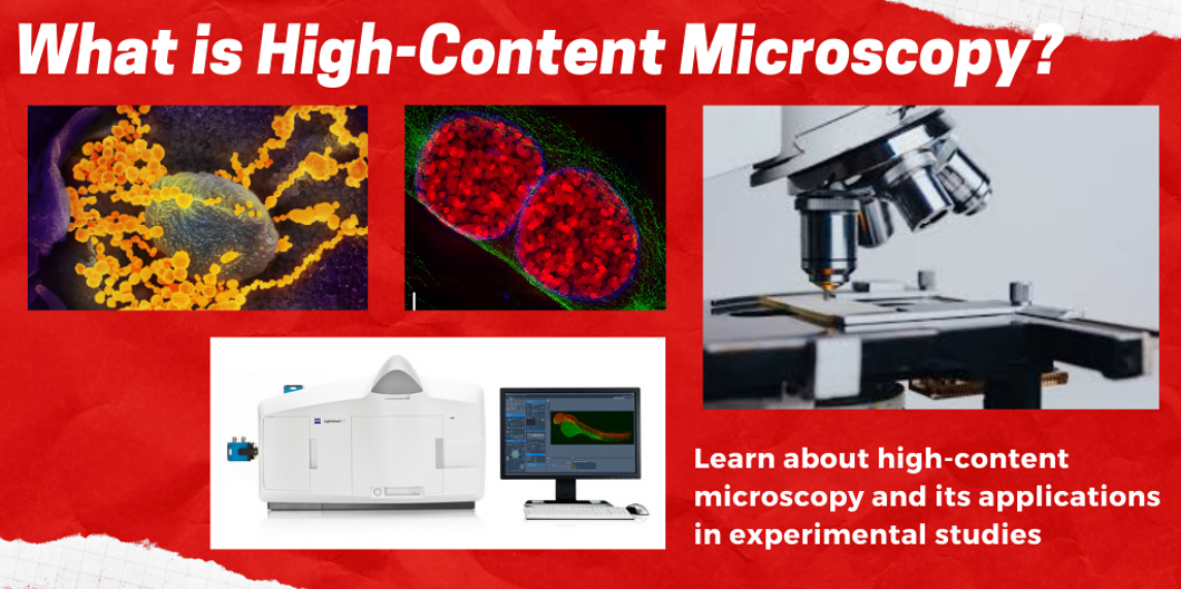
Figure 1. This lesson will introduce the technique of high-content microscopy and cover its advantages over traditional microscopy, its challenges and limitations, the steps for performing this technique, and its applications in experimental studies. The top left image shows the SARS-CoV-2 virus, the top middle shows a cell in prophase, the top right shows a traditional microscope, and the bottom image shows a high-content microscopy imager by Zeiss. Image created with Canva Pro.
Introduction
High-throughput technologies are slowly gaining traction and being implemented in many types of experiments, and most notably for drug discovery. An essential component of many scientific studies involves the use of microscopy. High-content microscopy is a tool that allows researchers to obtain vital information about the dynamics within cells1. These assays often require the use of fluorescent dyes to better visualize cells and their components. It is different from traditional microscopy in that more information can be obtained about the specimen being studied, and more complex cellular processes can be uncovered with this new, high-throughput approach to microscopy2. The resolution of high-content microscopy techniques have greatly surpassed that of traditional microscopy techniques, paving the way for more in-depth experimental studies3.
Advantages and Challenges
Advantages to high-content microscopy are the ability to study protein functions in single living cells, and to do this in a high-throughput way. Traditional microscopy methods often require the use of dyes to visualize the sample, which typically kills the microorganism being studied. They often cannot pinpoint a single cell for detailed images. With high-content microscopy, varying magnifications can be used to view both an entire population of microorganisms or the subcellular details of a single cell. Some high-content microscopes are able to emulate physiological conditions by adjusting the temperature, humidity, and CO2, which means the cells can be kept alive during the experimental process1.
A challenge that must be overcome with high-content microscopy lies in the image and data processing step. In an experiment that is studying many conditions, a large amount of images are taken for each condition. These images must all be processed, and often in the same way for consistency and better statistical analysis. Determining the best way to go about this step is a challenge that takes much time and thought to overcome1. Another challenge is the lack of advanced software to handle the data from the microscope. The software must be further developed to better handle the microscopic outputs at a high level1.
Steps for Conducting a HCM Study
To perform high-content microscopy there are five main steps that must be done. The sample is prepared, images are taken, data is collected and stored, the images are analyzed, and the data is modeled. These steps are often done in conjunction with other high-throughput methods like robotic liquid handlers for sample preparation1. Due to the vast amount of data that is produced with this technique, there is often advanced software in place to handle the output from the microscopic images and analyze the data.
Applications
There are many uses for high-content microscopy, but the most notable is drug discovery. With high-content microscopy, researchers are able to identify the precise molecular targets that a drug works on. The ability to “see” into the cellular processes will increase the number of potential drug targets that can be exploited which were not previously known with traditional microscopy1. Other applications include studying how cells behave and how they can be differentiated. This is particularly true when analyzing cells that make up a cancerous tumor, primary cell lines, recombinant cells, or stem cells2. A combination of high-content microscopy with techniques like RNAi, CRISPR, and functional genomics make these more efficient and precise because of the ability to visualize their exact mode of action within a cell4.
High-content microscopy is an extremely useful tool for researchers and will continue to increase our understanding of the world we live in. With the vast improvements and advancements in medicine it has proven itself time and time again to truly be more than just an image.
Resources
References
- Starkuviene, V. & Pepperkok, R. The potential of high-content high-throughput microscopy in drug discovery. Br. J. Pharmacol. 152, 62–71 (2005).
- Comley, J. Latest developments in high content screening systems. https://www.ddw-online.com/screening/p315004-latest-developments-in-high-content-screening-systems.html. (2016).
- Pepperkok, R. & Ellenberg, J. High-throughput fluorescence microscopy for systems biology. Nat. Rev. Mol. Cell Biol. 7, 690–696 (2006).
- Kroll, T., Schmidt, D., Schwanitz, G., Ahmad, M., Hamann, J., Schlosser, C., Lin, Y. C., Bohm, K. J., Tuckermann, J., & Ploubidou, A. High-content microscopy analysis of subcellular structures: assay development and application to focal adhesion quantification. Curr. Protoc. Cytom. 77, 12.43.1-12.43.44 (2016).
Questions
- What is high-content microscopy? This is a high-throughput imaging method that can provide sub-cellular details about a single living cell to observe in more detail the dynamics of the interactions that occur within a cell.
- Why is it important? High-content microscopy generates lots of data in an efficient way. It allows for a large amount of information to be gathered about a single cell or population of cells, which can more efficiently progress the knowledge on a particular subject (protein-protein interactions, drug interactions, biochemical processes..etc.). It can streamline the experimental process as many conditions can be created and observed using multi-well plates or imagers that can control environmental conditions.
- List the steps a researcher would need to take to perform a high-content microscopy study. Sample preparation, image acquisition, storage and collection of the images, analysis of the images, and modeling of the data obtained throughout the process.
- Why might a researcher choose to use high-content microscopy over traditional microscopy? High-content microscopy will allow the researcher to study the cell/process in more detail. The resolution of this technique is better and can provide more information about the dynamics within cells.